Prostate cancer is a leading cause of death among men, and a disease severely diminishing quality of life with complications of
sexual, urinary, and rectal dysfunction.
Our goal, with our collobartors from University of Toronto, is to develop automated cancer localization methods based on the reliable ground truth that are obtained from histology and multispectral MRI.
The methods that we develop will be used to guide biopsy, radiotherapy and even possibly surgery.
Centers performing routine radical prostatectomy could alter their surgical approach to ensure negative margins if they had access to pre-operative knowledge of tumor volume and localization. Results of this effort could also open the way to subtotal-ablative or dose-escalation approaches to therapy, minimizing complications while maximizing efficacy. If a reliable method were available to acquire and automatically analyze multispectral MRI data to localize cancer, it would allow for optimized targeting of TRUS biopsy, and improved surveillance of small indolent cancers, potentially leading to a reduction in mortality through patient-tailored therapy.
Automated 3-D segmentation of the prostate and the tumor. Left: Ground truth, right: our automated methods.
Green color shows the surrounding tissue and organs, blue the prostate, and red the tumor.
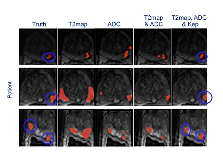
Using Multispectral MRI increases segmentation accuracy. From left to right: increased number of
MR image types. Ground truth shown in the first column is obtained from pathalogy.
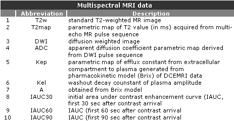
Description of MR image types.
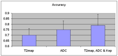
Quantitative results. Accuracy increases with increased number of MR image types.
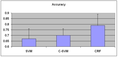
Advanced segmentation methods we have developed, such as cost-sensitive SVM and CRF (conditional random fields),
significantly increases segmentation accuracy. For this figure, the images T2map, ADC and Kep are used.
We have also developed an unsupervised segmentation algorithm based on fuzzy Markov random fields and found that
increasing number of types of images increase the performance segmentation here as well. We have shown that the method
we have developed is superior to classical unsupervised segmentation methods. Please see references below
for more details.
In another study, schemes for threshold selection for both supervised and unsupervised algorithms, and shown
that we can improve segmentation accuracy by such threshold selection methods. We have also compared the
supervised and unsupervised techniques and found that supervised algorithms are superior to unsupervised methods.
Please see references below for more details.
References:
X. Liu, M. A. Haider, D. L. Langer, I. S. Yetik, "A Tumor Segmentation Method Based on Local Contrast for Improved
Localization Performance", submitted.
L. Liu, M. A. Haider, D. L. Langer, I. S. Yetik, "Prostate Cancer Localization with Spatially Regularized Parametric Images Obtained
from DCE MRI", in revision, IEEE Trans. on Image Processing.
Y. Artan, M. A. Haider, D. L. Langer, T. H. van der Kwast, A. J. Evans, Y. Yang, M. N. Wernick, J. Trachtenberg,
I. S. Yetik, "Prostate Cancer Localization with Multispectral MRI Using Cost-Sensitive Support Vector Machines and
Conditional Random Fields", to appear, IEEE Transactions on Image
Processing .
Y. Artan, M. A. Haider, D. L. Langer, I. S. Yetik,"Semi-Supervised Prostate Cancer Segmentation with
Multispectral MRI," Proceedings of IEEE International Symposium on Biomedical Imaging, pp. 648-651, 2010.
L. Liu, M. A. Haider, D. L. Langer, I. S. Yetik, "Improved Prostate Cancer Localizaiton with Spatially
Regularized Dynamic Contrast-Enhanced Magnetic Resonance Imaging," Proceedings of IEEE International Symposium on Biomedical
Imaging, pp. 644-647, 2010.
X. Liu, M. A. Haider, D. L. Langer, I. S. Yetik, "Using Relative Contrast and Iterative Normalization for
Improved Prostate Cancer Localizaiton with Multispectra MRI", Proceedings of IEEE International Symposium on Biomedical
Imaging, pp. 1369-1372, 2010.
S. Ozer, D. L. Langer, X. Liu, M. Haider, T. H. van der Kwast, A. J. Evans, Y. Yang, M. N. Wernick, I. S.
Yetik, "Supervised and Unsupervised Methods for Prostate Cancer Localization with Multispectral MRI",
Medical Physics, pp. 1873-1883, 2010.
X. Liu, D. Leager, M. Haider, M. Wernick, Y. Yang, I. S, Yetik, "Prostate Cancer Segmentation with
Simultaneous Estimation of Markov Random Field Parameters and Classes", IEEE Transactions on Medical Imaging,
vo. 28, no. 6, pp. 906-915, 2009.
S. Ozer, M. Haider, D. Langer, T. H. van der Kwast, A. J. Evans, M. Wernick, J. Trachtenberg, I. S. Yetik, "Prostate Cancer Localization with
Multispectral MRI Based on Relevance Vector Machine", Proceedings of IEEE International Symposium on Biomedical Imaging, pp. 73-76, 2009.
Y. Artan, D. Langer, M. Haider, T. H. van der Kwast, A. J. Evans, M. N. Wernick, I. S. Yetik, "Prostate Cancer Localization with Multispectral
MRI Using Conditional Random Fields", Proceedings of IEEE InternationalSymposium on Biomedical Imaging, pp. 282-285, 2009.
X. Liu, I. S. Yetik, M. N. Wernick, "Simultaneous estimation of the Markov Random Field parameters and the classes for image segmentation",
Proceedings of IEEE International Conference on Image Processing,pp. 3048-3041, 2008.