Oxygen Tension Imaging of Rat Retina
Abnormalities in retinal oxygenation are thought to play a significant role in the development of common eye diseases,
such as diabetic retinopathy, glaucoma, and age-related macular degeneration.
However, our knowledge of oxygen’s function in many retinal diseases is incomplete.
Therefore, accurate assessment of oxygen tension in the retinal vasculature is important to
establish the role of oxygen in the development of retinal disease and to aid in the detection of the onset or presence of retinal diseases.
Our goal, with a team from University of Illinois at Chicago, is to develop advanced image reconstruction schemes to obtain more accurate oxygen maps.
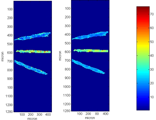
Reconstruction methods we develop based on spatial regularization significantly
improves the quality of the oxygen maps.
Left: Standard reconstruction, right:
the reconstruction method we have developed.
The data is collected from rat retina.
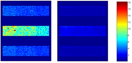
Comparison of the standard deviation of estimated images.
Left: Standard reconstruction, right:
the reconstruction method we have developed.
We observe that the variance of reconstructed
image is significantly lower when our method is used.
References:
- I. Yildirim, R. Ansari, J. Wanek, I. S. Yetik, M. Shahidi, "Regularized Estimation of Retinal Vascular Oxgen Tension from Phosphorescence
Images", IEEE Transactions on Biomedical Engineering, vol. 56, pp. 1989-1995, 2009.
- I. Yildirim, R. Ansari, J. Wanek, I. S. Yetik, M. Shahidi, "Denoising Algorithms and Retinal Oxygen Tension Estimation with EM",
Proceedings of IEEE International Symposium on Biomedical Imaging, pp. 57-60, 2009.
- I. Yildirim, R. Ansari, J. Wanek, I. S. Yetik, M. Shahidi, "Retinal oxygen tension estimation in phosphorescence lifetime images using
regularized least squares", Proceedings of IEEE Electro/Information Technology, pp. 465-469, 2008.
|